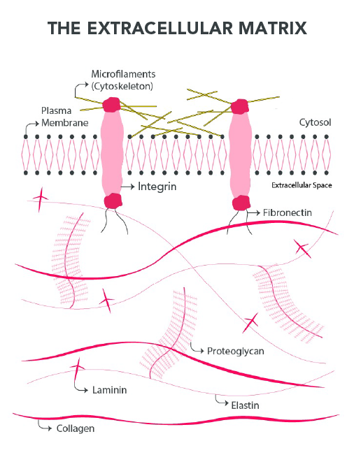
The extracellular matrix (ECM) is a highly complex and dynamic three-dimensional structure present in all tissues and organs. It provides vital biochemical and biomechanical clues necessary for tissue morphogenesis, differentiation, and homeostasis. It also plays a crucial role in the structural and functional integrity of organs and tissues, acting as a support for cell attachment. The ECM is mainly composed of large and insoluble structural proteins, glycosaminoglycans (GAGs), collagen, and elastin, that interact with each other forming an intricate network [1,2].
Dysregulation of ECM composition and structure is associated with the development and progression of several pathologic conditions. Diseases such as osteogenesis imperfecta - a congenital disease that primarily causes low bone mass and bone fractures - are characterized by alterations in quantity, quality, mineralization, and homeostasis of the extracellular matrix [3]. In addition, several pathological conditions, such as fibrosis and invasive cancer, are also related to dysregulation of ECM structure, stiffness, composition, and abundance [4].

Because of the important role of the ECM in the structure and function of tissue and organs, many researchers believe bioprinting decellularized ECM (dECM) is a promising strategy and a potential choice for regenerative scaffold and tissue fabrication. Decellularized extracellular matrix-based bioinks can offer an optimized microenvironment for the growth of 3D structured tissues, as they provide specific biological and mechanical cues from the original ECM and the right proportions of ECM proteins, mimicking the complex microenvironment found in the native tissue by cells [5,6].
Pati et al. (2014) studied the decellularization of cartilage, adipose, and heart tissues to develop dECM bioinks capable of providing a complex and specific microenvironment for stem cell growth. The researchers developed an optimized process to remove the cellular content of the tissues while keeping important ECM proteins, which was demonstrated by the quantification of the content of GAGs and collagen in the decellularized material. The rheological behavior of the dECM bioinks was also evaluated and all formulations presented shear-thinning behavior and gelation above 15ºC, which is desirable for their application in extrusion-based bioprinting. 3D bioprinted dECM-based constructs were produced either by depositing the cell-laden dECM pre-gel alone or following fabricating of PCL framework. The printed cell-laden constructs presented higher levels of cell viability, differential lineage commitment and ECM formation. This cell-laden dECM hydrogel deposition technology fulfilled several vital requirements of tissue engineering grafts, such as the introduction of pertinent cells and the opportunity to explore organizational aspects in tissue regeneration [6].

Figure 2: Schematic representation of 3D bioprinting using dECM bioink encapsulating living stem cells with a polymeric framework and possible applications [6].
As an alternative to liver transplantation, Mao et al. (2020) developed a dECM-based cell-laden bioink for liver microtissue fabrication. The bioink was created by combining a photocurable material, GelMA (gelatin methacrylate), with dECM and human-induced hepatocytes (hiHep cells). Mao and his group used the light-based technology (digital light processing, DPL) to fabricate 3D bioprinted constructs. The researchers reported that the liver dECM-based bioink showed an excellent printability, high viability of hiHep cells, and the liver microtissue expressed excellent liver functions [7].
These works demonstrate the possibility of developing dECM-based bioinks suitable for different 3D bioprinting technologies and the importance of these materials to develop complex 3D constructs able to recreate the microenvironmental characteristics of different tissues and organs, truly representing their native ECM.
Want to know more about the concepts of the ECM, its composition, role in the stem cell niche, and other important information about this vital component of living tissues? Check our Ebook "Extracellular matrix - Composition, Organization, and Regulation of Stem Cell Behavior".
REFERENCES
1. Frantz, C., Stewart, K. M. & Weaver, V. M. The extracellular matrix at a glance. J. Cell Sci. 123, 4195–4200 (2010).
2. Kusindarta, D. L. & Wihadmadyatami, H. The Role of Extracellular Matrix in Tissue Regeneration. in Tissue Regeneration (ed. hay El-Sayed Kaoud, H. A.) (IntechOpen, 2018).
4. Bonnans, C., Chou, J. & Werb, Z. Remodelling the extracellular matrix in development and disease. Nat. Rev. Mol. Cell Biol. 15, 786–801 (2014).
5. Dzobo, K., Motaung, K. S. C. M. & Adesida, A. Recent Trends in Decellularized Extracellular Matrix Bioinks for 3D Printing: An Updated Review. Int. J. Mol. Sci. 20, (2019).
6. Pati, F. et al. Printing three-dimensional tissue analogues with decellularized extracellular matrix bioink. Nat. Commun. 5, 3935 (2014).
7. Mao, Q. et al. Fabrication of liver microtissue with liver decellularized extracellular matrix (dECM) bioink by digital light processing (DLP) bioprinting. Mater. Sci. Eng. C Mater. Biol. Appl. 109, 110625 (2020).

Commentaires